
By Dr. Muhammad Awais Rabbani, MBBS · Medically reviewed by Dr. Ahmed Humayon, MBBS · Updated 12 November 2026 · Editorial policy
RDW (red cell distribution width) measures how much your red blood cells vary in size. A high RDW means the sizes vary more than usual, often from low iron, vitamin B12 or folate; doctors read it alongside MCV.
Already have your report? Enter your RDW and MCV below.
Check your RDW result
Enter the RDW from your report, pick RDW-CV (%) or RDW-SD (fL), and keep or replace the reference interval printed beside it. Add your MCV, and the RDW result checker shows which RDW–MCV pattern your RDW results fit, what that pattern is commonly associated with, and the tests a doctor usually orders next.
Ranges: Mayo Clinic Laboratories; Brill & Baumgardner 2000. Patterns: Bessman 1983. This does not diagnose anything.
The checker uses the range on your own report first, because RDW ranges differ from lab to lab. If you leave the boxes as they are, it falls back to published adult defaults: RDW-CV 11.8–14.5% for men and 12.2–16.1% for women, and MCV 78.2–97.9 fL, from Mayo Clinic Laboratories, plus RDW-SD 39–47 fL from a reference in American Family Physician.
It answers three things in order. First, whether your RDW sits below, inside or above the range. Second, if you added MCV, which of six patterns the two values form. Third, which follow-up tests usually come next for that pattern. Hemoglobin is a simple low or normal choice on purpose, because the checker reads patterns and never names a diagnosis.
When both RDW and MCV sit inside your ranges, it says so and stops there. The six-pattern grid was built to sort anemias, so it means most when hemoglobin is low.
Tool sources: Mayo Clinic Laboratories (RDW-CV, MCV); Brill and Baumgardner, 2000 (RDW-SD); Bessman and colleagues, 1983, as reproduced in Rodak’s Hematology (pattern grid). Tool logic and ranges reviewed by Dr. Ahmed Humayon, MBBS. Not a diagnosis.
What is RDW in a blood test?
RDW, short for red cell distribution width, is a value on your complete blood count (CBC) that shows how much the size of your red blood cells varies from one cell to the next. A low spread means the cells are close to uniform. A wide spread means a mix of small and large cells, a finding doctors call anisocytosis.
Some reports print the full name, red blood cell distribution width.
Red blood cells carry oxygen from your lungs to the rest of your body, and each one lives about 120 days before the bone marrow replaces it. The marrow releases healthy red blood cells at a fairly even size. When the marrow runs short of iron, vitamin B12 or folate, or when cells are lost or destroyed faster than usual, new cells come out a different size from the older ones still circulating, and the mix shows up as a wider spread.
The RDW measures that size variation. It does not say which direction the sizes have moved. That job belongs to the mean corpuscular volume (MCV), which reports the average size of the same cells.
MCV gives the average. RDW gives the spread around it.
That is why RDW is read with MCV and hemoglobin, never alone. A wide spread around a small average means something different from a wide spread around a large one, and the same RDW can sit beside a normal or a low hemoglobin.
What is a normal RDW range?
A normal RDW is the range printed next to the result on your own lab report. RDW-CV is reported as a percentage and RDW-SD in femtoliters (fL), and each has its own reference range. MedlinePlus notes that the range varies by lab, so the published defaults below are a fallback for when your report shows none.
| Measure | Unit | Published adult reference interval | Source |
|---|---|---|---|
| RDW-CV | % | Men 11.8–14.5; women 12.2–16.1 | Mayo Clinic Laboratories, CBC test, adults 18 and over |
| RDW-SD | fL | 39–47 | Brill and Baumgardner, American Family Physician, 2000 |
| MCV | fL | 78.2–97.9 | Mayo Clinic Laboratories, CBC test, adults 18 and over |
Your lab’s range wins. If a website table and your report disagree, use the range on your report. It was set for the analyzer and the population that produced your number.
RDW is calculated by the hematology analyzer from the red cell size curve, so a result above the normal range on one lab’s report can sit inside it on another’s. Even one lab’s range can shift with the person tested. Mayo prints different RDW-CV limits for men and women, and a study of 8,089 people found that RDW rose with age. If your number looks high against a table online but falls inside the range on your report, your report is the one your doctor reads.
A letter H or L beside the result means it fell outside the lab’s interval.
A normal RDW on its own does not rule anything in or out.
What does a high RDW mean?
A high RDW means your red blood cells vary in size more than your lab’s range allows. It reflects a mix of older and newer cells of different sizes. Common causes are nutritional deficiencies, mainly low iron, vitamin B12 or folate, along with blood loss and hemolysis. A high RDW alone is not a diagnosis.
An elevated RDW can show up early. In iron deficiency, a wider spread of cell sizes may be the first abnormal lab finding, appearing before anemia and before the cells turn small. That is early iron deficiency: the marrow starts releasing smaller cells while older, normal-sized cells still circulate, so the spread widens before the average moves.
What a high RDW points to depends on the MCV beside it. A high RDW with a low MCV suggests one group of causes, a high RDW with a high MCV another, and a high RDW with a normal MCV a third. The section on reading RDW with MCV sets out all six combinations, and the causes list explains each one.
RDW also moves slowly.
Red cells last about 120 days, so a change in the cells being made takes weeks to show across the whole population. The same delay works in reverse. Early in iron treatment, the RDW can rise as new normal-sized cells join the small ones. A rising RDW during treatment is therefore not always a bad sign, and your doctor reads it against the hemoglobin trend.
How far above the range a result sits matters less than what sits next to it on the report.
What does a low RDW mean?
A low RDW means your red blood cells are more uniform in size than your lab’s range expects. MedlinePlus says a low result is not a sign of anemia and usually nothing to worry about, and no disease is known to cause a low RDW. Doctors look at the rest of the CBC instead.
People often search for low RDW symptoms. A low RDW does not cause symptoms by itself. If you feel tired or unwell and your report also shows a low RDW, the RDW is unlikely to be the reason; something else on the report, or something outside the blood count, has to explain it. Hemoglobin, MCV, the white cell count and the platelet count, including platelet size (MPV), are where a doctor looks next.
A low result is worth raising with your doctor when:
- it sits next to other flagged values, such as low hemoglobin or a high or low MCV
- you had symptoms that led to the test in the first place
- earlier reports showed a different pattern and you want to know why it changed
Because a low RDW is not clinically significant on its own, the checker above reports it without a follow-up list. It still shows the hemoglobin note if you marked hemoglobin as low, and it still applies your lab’s own lower limit rather than a website default.
RDW-SD vs RDW-CV: what’s the difference?
RDW-CV and RDW-SD are two ways of measuring the same spread. RDW-CV divides the standard deviation of red cell volume by the MCV and gives a percentage, so it depends on MCV. RDW-SD measures the width of the size curve in femtoliters, at 20% of its height, so MCV does not affect it. Many reports print only one of them.
| RDW-CV | RDW-SD | |
|---|---|---|
| What it measures | Spread of cell size relative to the average (MCV) | Width of the red cell size curve itself |
| Unit | % | fL |
| Calculated as | (standard deviation of cell volume ÷ MCV) × 100 | Curve width at 20% of its height |
| Affected by MCV? | Yes, because it is divided by MCV | No |
| What “high” means | More size variation than the lab’s % range | More size variation than the lab’s fL range |
Picture two samples whose cells spread across the same range of sizes. If one sample has a smaller average cell, dividing by that smaller MCV makes its RDW-CV higher, while its RDW-SD stays the same. With a larger average cell, the opposite happens. That is the whole reason the two numbers can disagree on the same tube of blood.
Age shows the same split. In the 8,089-person study, conventional RDW rose about 6% from the youngest to the oldest age group while RDW-SD rose nearly 15%, a gap the authors traced to MCV climbing with age.
If your report shows only one of them, that is normal. Read the one you have against its own reference range. The two use different units, so an RDW-CV figure and an RDW-SD figure can never be compared directly. In the checker above, pick the unit that matches your report.
A report that just says “RDW” with a percent sign is giving you RDW-CV.
What causes a high RDW?
The common causes of a high RDW are iron deficiency, vitamin B12 deficiency and folate deficiency. Each makes the marrow release red cells of a different size from the ones already circulating. Recent blood loss, hemolysis, a recent blood transfusion, liver disease, kidney disease and chronic inflammation can raise it too.
- Iron deficiency: new cells come out smaller, and iron deficiency anemia is the classic high-RDW, low-MCV picture.
- Vitamin B12 deficiency: new cells come out larger than the ones they replace.
- Folate deficiency: same direction as B12, toward larger cells.
- Mixed deficiency: iron plus B12 or folate can leave two populations of cells, called a dimorphic picture.
- Recent blood loss: the marrow speeds up and releases young cells while iron stores run down.
- Hemolysis: when red cells break down early, the marrow releases young cells that are larger than mature ones; a bilirubin blood test is one of the checks for it.
- Recent blood transfusion: donor cells mix with your own for a while, which widens the spread.
- Liver disease: chronic liver disease and alcohol-related anemia both raise it.
- Kidney disease: anemia is common in long-term kidney disease, which is checked with kidney function on the same panel of blood tests.
- Chronic inflammation: long-standing inflammation affects iron use; your doctor may also check the ESR, another marker of inflammation.
- Bone marrow disorders: myelodysplastic syndrome can produce cells of uneven size.
- Cancer: MedlinePlus lists cancer, especially colorectal cancer, though a high RDW is not a cancer test.
Two causes can run at once.
Iron deficiency pulls the average cell size down while B12 or folate deficiency pushes it up, so MCV can land in the normal range even though the RDW is high. That is the dimorphic picture, and it is one reason a normal MCV does not close the question when RDW is raised. It is also why the follow-up tests below check iron, B12 and folate together instead of one at a time.
How doctors read RDW with MCV
Doctors read RDW together with MCV, the average size of your red cells. MCV says whether the cells are small (microcytic), normal (normocytic) or large (macrocytic); RDW says whether they are uniform or mixed. The six combinations narrow the list of likely causes of anemia. This scheme is the Bessman classification, published in 1983.
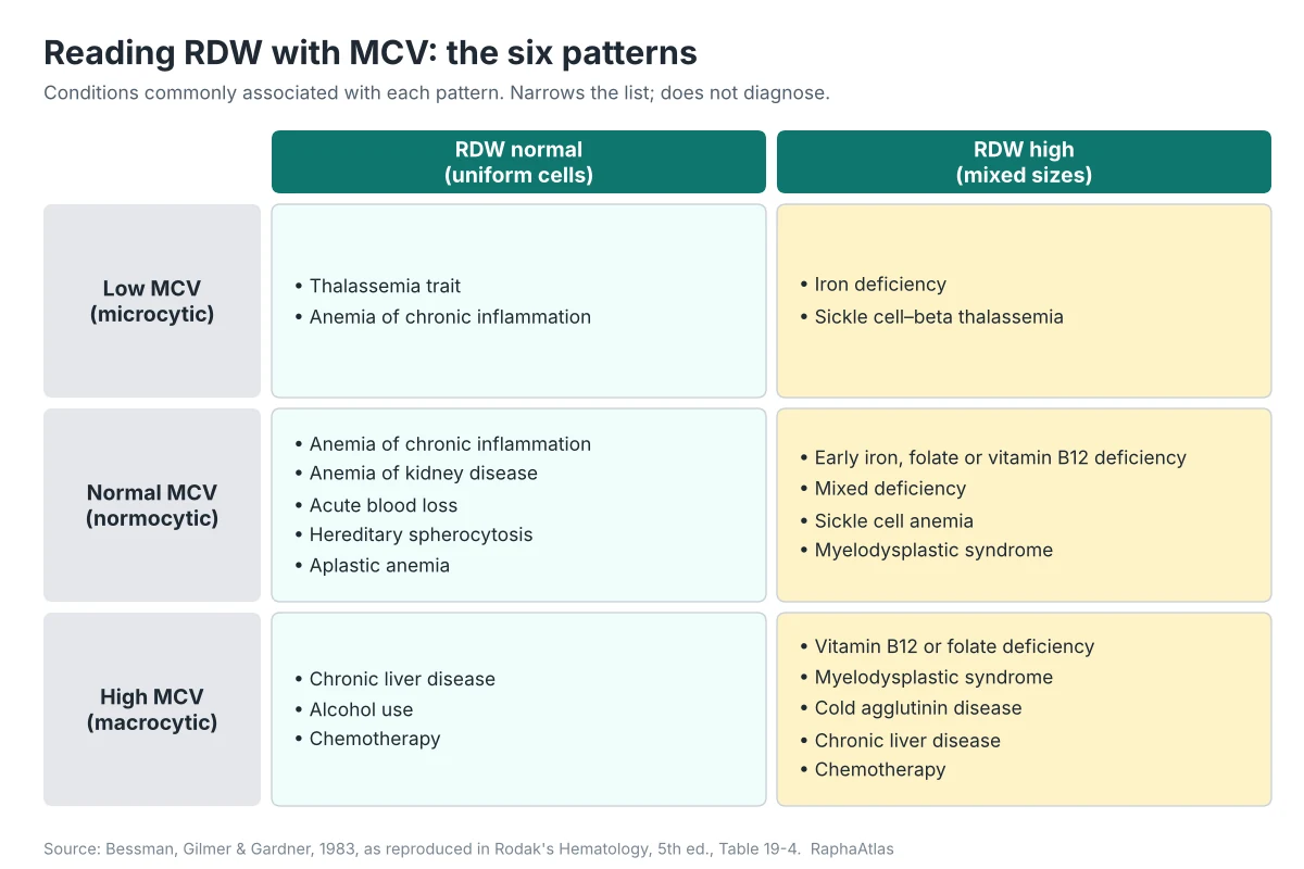
| MCV | RDW normal | RDW high |
|---|---|---|
| Low (microcytic) | Thalassemia trait; anemia of chronic inflammation | Iron deficiency; sickle cell–beta thalassemia |
| Normal (normocytic) | Anemia of chronic inflammation; anemia of kidney disease; acute blood loss; hereditary spherocytosis; aplastic anemia | Early iron, folate or vitamin B12 deficiency; mixed deficiency; sickle cell anemia; myelodysplastic syndrome |
| High (macrocytic) | Chronic liver disease; alcohol use; chemotherapy | Vitamin B12 or folate deficiency; myelodysplastic syndrome; cold agglutinin disease; chronic liver disease; chemotherapy |
Grid: Bessman, Gilmer and Gardner, 1983, as reproduced in Rodak’s Hematology, Table 19-4. The textbook notes the scheme is not absolute, because RDW values overlap between conditions.
Take a CBC with low hemoglobin, low MCV, low MCH and a high RDW. That combination fits the microcytic, high-RDW cell, and iron deficiency sits at the top of that cell’s list. A low mean corpuscular hemoglobin (MCH) points the same way, because small cells made without enough iron also carry less hemoglobin. The same low MCV with a normal RDW reads differently: in beta thalassemia trait the cells are uniformly small, and the RDW tends to stay normal.
These RBC indices work as a set.
The macrocytic row splits too. Large cells with a high RDW lean toward vitamin B12 or folate deficiency or myelodysplastic syndrome. Large, uniform cells lean toward chronic liver disease, alcohol use or chemotherapy. In both rows, B12 and folate are usually checked early, because those deficiencies are common and treatable.
The middle row is where RDW earns its place. A normal MCV with a high RDW is where early iron, B12 or folate deficiency and mixed deficiency sit, before the average cell size has moved or while two changes cancel out.
The grid narrows the list. It does not diagnose. Each cell holds several conditions, and real results often sit near a border. Ferritin, B12, folate, a reticulocyte count and a blood smear decide which one applies, as the next sections show.
Cut-offs for low, normal and high MCV come from your lab’s reference interval as well.
Is RDW part of a CBC?
Yes. RDW is part of the complete blood count (CBC), and MedlinePlus describes the RDW test as often run within a CBC. The analyzer calculates it from the same blood sample used for hemoglobin, MCV, the red blood cell count and the other red cell values. All of them come from that one sample, so RDW needs no tube of its own.
One blood draw covers the whole panel.
The analyzer builds a curve of red cell sizes from that sample, reports MCV as its average and RDW from its width. No extra tube, fasting or preparation is needed for RDW beyond what the CBC itself requires. If a doctor wants RDW rechecked, the request is simply a repeat CBC.
From the billing side. The CBC is one line on a claim. In Medicare’s national lab data, CPT 85025 is the complete CBC with automated differential white cell count, and CPT 85027 is the complete automated CBC without it. RDW is reported inside either test, so it does not appear as its own charge on a bill or Explanation of Benefits.
What happens after a high RDW result?
After a high RDW, a doctor usually looks at hemoglobin and MCV first, then orders tests to find the cause. The usual next tests are ferritin and iron studies, vitamin B12, folate, a reticulocyte count and a peripheral blood smear, sometimes with a repeat CBC. A primary-care review names RDW, a reticulocyte index and the smear as the basic first studies.
- Ferritin and iron studies: ferritin is the usual first test for iron stores.
- Vitamin B12: checked when cells run large or the picture looks mixed.
- Folate: checked alongside B12 for the same reason.
- Reticulocyte count: counts young red cells and shows whether the bone marrow is responding to blood loss or hemolysis.
- Peripheral blood smear: a trained person looks at your cells under a microscope and can see a dimorphic population directly.
- Repeat CBC: confirms the result and shows the trend.
An isolated high RDW, meaning a high RDW with normal hemoglobin and a normal MCV, fits the early-deficiency cell of the grid. That is why ferritin, B12 and folate are reasonable checks even when nothing else on the CBC is flagged. If a repeat is back in range and you feel well, it often needs nothing more. If it stays high, the tests above help determine the cause.
If your hemoglobin is also low, ask how soon the follow-up should happen.
Take the result to your health care provider with any earlier test results you have. Two or three reports in a row say more than one number, and an RDW that has been stable for years reads very differently from one that rose since your last CBC.
Why doctors pay attention to RDW beyond anemia
Researchers have linked a higher RDW with worse outcomes in heart disease, which is why it is studied as a prognostic marker. A 2012 review reported associations with heart attack, heart failure, stable angina, stroke and peripheral artery disease. These are associations across groups of patients. RDW is not a test for heart disease or any other illness.
The same review called the reasons not fully understood. Proposed explanations include chronic inflammation, oxidative stress and reduced kidney function, which can affect red cells and blood vessels at the same time. Other studies extend the link, including one tying high RDW to carotid artery atherosclerosis in people with high blood pressure, and MedlinePlus lists heart disease and diabetes among conditions seen with a high RDW.
A higher RDW across a study population does not mean RDW caused the outcome, and it cannot predict what will happen to one person. Doctors do not order a CBC to screen for heart disease through the RDW. If you have a heart condition and your RDW is raised, ask your doctor whether it changes anything in your care, with the full CBC in front of you. Most of the time the answer comes back to the question behind anyone else’s result: what is causing it?
For your own result, the practical next step is still the follow-up workup above.
RDW blood test FAQ
What vitamin deficiency causes high RDW?
Vitamin B12 deficiency and folate deficiency both raise RDW, usually with a high MCV. Iron deficiency raises it too, usually with a low MCV, though iron is a mineral rather than a vitamin. The causes section covers the rest.
Is RDW high in pregnancy?
It can be, but studies disagree. A 2018 systematic review found one large study where RDW-CV upper limits rose across the three trimesters and another where RDW did not change. RDW alone is not a reliable iron test in pregnancy, so your prenatal team reads it with hemoglobin, MCV and ferritin.
Can RDW be high when MCV is normal?
Yes. A high RDW with a normal MCV fits early iron, B12 or folate deficiency, or a mixed deficiency where small and large cells average out. It is the pattern where the RDW tells a doctor more than the MCV does, and ferritin, B12 and folate usually come next. See the RDW and MCV grid.
What does RDW stand for?
RDW stands for red cell distribution width. Some reports spell it out as red blood cell distribution width, or label it RDW-CV or RDW-SD, depending on how the analyzer expresses the spread of cell sizes.
Can a high RDW go back to normal?
It can, but slowly. Red cells live about 120 days, so the mix of sizes changes over months once the cause is treated, and RDW can even rise early in iron treatment. A repeat CBC shows the trend.
How does a doctor read an RDW result?
Hemoglobin first, to see whether there is anemia at all. Then MCV, to place the cells as small, normal or large. Then the RDW against the lab’s own range, and last any earlier CBC, because a change over time says more than one value.
Sources
- MedlinePlus. RDW (Red Cell Distribution Width). U.S. National Library of Medicine. https://medlineplus.gov/lab-tests/rdw-red-cell-distribution-width/
- Mayo Clinic Laboratories. Complete Blood Cell Count (CBC) with Differential, Blood (Test ID: CBC), reference values. https://www.mayocliniclabs.com/test-catalog/Overview/CBC
- Brill JR, Baumgardner DJ. Normocytic anemia. Am Fam Physician. 2000;62(10):2255–2263. https://www.aafp.org/afp/2000/1115/p2255
- Bessman JD, Gilmer PR Jr, Gardner FH. Improved classification of anemias by MCV and RDW. Am J Clin Pathol. 1983;80(3):322–326. PMID: 6881096. Grid as reproduced in: Keohane EM, Smith LJ, Walenga JM, eds. Rodak’s Hematology: Clinical Principles and Applications. 5th ed. Elsevier; 2016. Chapter 19, Table 19-4.
- Sarma PR. Red Cell Indices. In: Walker HK, Hall WD, Hurst JW, eds. Clinical Methods. 3rd ed. Butterworths; 1990. Chapter 152. https://www.ncbi.nlm.nih.gov/books/NBK260/
- Aird W. Red cell distribution width (RDW). The Blood Project; updated 10 November 2025. https://www.thebloodproject.com/red-cell-distribution-width-rdw-2/
- Effect of age and gender on reference intervals of red blood cell distribution width (RDW) and mean red cell volume (MCV). Clin Chem Lab Med. 2015. doi:10.1515/cclm-2015-0155
- Isik T, Ayhan E, Kurt M, Tanboga IH, Kaya A, Aksakal E. Is red cell distribution width a marker for the presence and poor prognosis of cardiovascular disease? Eurasian J Med. 2012;44(3):169–171. doi:10.5152/eajm.2012.39
- High red blood cell distribution width is closely associated with risk of carotid artery atherosclerosis in patients with hypertension. PMC2954027. https://pmc.ncbi.nlm.nih.gov/articles/PMC2954027/
- Paliogiannis P, Zinellu A, Mangoni AA, et al. Red blood cell distribution width in pregnancy: a systematic review. Biochem Med (Zagreb). 2018;28(3):030502. doi:10.11613/BM.2018.030502
- Centers for Medicare & Medicaid Services. Medicare Part B Physician/Supplier National Data CY2019: Top 100 Lab Procedures (85025, 85027). https://www.cms.gov/research-statistics-data-and-systems/statistics-trends-and-reports/medicarefeeforsvcpartsab/downloads/labserv19.pdf
- Stanford Blood Center. How long do red blood cells live? https://stanfordbloodcenter.org/how_long_do_red/